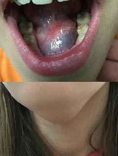
(BMJ)—A 12-yo girl presented w/ a 3-mo hx of a mass in the floor of her mouth and swelling in the submental area. She had difficulty chewing and swallowing solids. Exam: nontender, fluctuant, bluish mass on L side of floor of mouth. No lymphadenopathy. Nontender external midline swelling. MRI confirmed the dx. What is it?
Thyroglossal duct cyst
Cystic hygroma
Plunging ranula
Cervical thymic cyst
Branchial cleft cyst
You are correct. MRI showed an hourglass-shaped cystic mass that extended from the oral cavity to the submental region and was filled w/ homogenous fluid, suggestive of plunging ranula. A ranula is a mucocele of the floor of the mouth, arising from an isolated accessory salivary gland or the sublingual gland. It’s classified as simple or plunging. Simple ranula is much more common and is confined to the oral cavity. Plunging ranula extends through the fibers of the mylohyoid muscle in the anterior neck, from the sublingual space into the submandibular space. Standard tx includes surgical removal of the ipsilateral sublingual gland and the associated ranula. Both parts of the lesion were resected transorally in this pt.

Arch Dis Child 2020;105:512