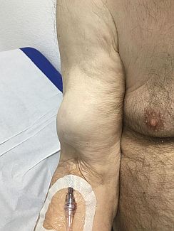
(BMJ)—An 83-yo man w/ DM, hypertension, chronic kidney dz, and asthma was admitted for community-acquired pneumonia. Home meds: candesartan, gliclazide, metformin, simvastatin, inhaled fluticasone/salmeterol. He improved on IV levofloxacin and ceftriaxone, but he experienced sudden right arm pain and swelling on day 4. What is the dx?
Extravasation injury
Bicipitoradial bursitis
Proximal biceps avulsion
Brachial artery aneurysm
Morel-Lavallee lesion
You are correct. Exam revealed a nonpulsatile, palpable mass in the mid-upper arm corresponding to the belly of the biceps muscle, consistent w/ proximal biceps avulsion. U/S confirmed the dx. The IV site showed no signs of phlebitis or extravasation. Absence of trauma ruled out bicipitoradial bursitis and Morel-Lavallee lesion. In most cases, a biceps brachii rupture occurs at the proximal insertion, involving the long head of the muscle. It is more common in the elderly w/ hx of chronic shoulder pain, recurrent microtrauma, or flexion against forced extension. Although rare, spontaneous tendon ruptures are assoc w/ DM, fluoroquinolones, statins, corticosteroids, end-stage kidney dz, hypothyroidism, hyperparathyroidism, connective tissue dz, and gout, several of which were present in this pt.

Emerg Med J 2019;36:153-162.
(BMJ)—A 27-yo woman presented w/ a 7-yr hx of a progressively worsening, edematous plaque over her L eyelid that was aggravated by sunlight. Review of sx: no joint pain/muscle weakness/mouth ulcers/hand lesions. Exam: visual acuity and eye movement normal. Labs: raised LDH. What is the dx?
Polymorphous light eruption
Periorbital cellulitis
Amyopathic dermatomyositis
Discoid lupus erythematosus
Cavernous sinus thrombosis
You are correct. A clinical dx of amyopathic dermatomyositis was made based on eye lesion w/ heliotrope rash and raised LDH, LDH1, and α-hydroxybutyrate dehydrogenase but no muscle weakness. Absence of joint pain, muscle weakness, mouth ulcers, and hand pathology made systemic lupus erythematosus or classic dermatomyositis unlikely. This pt’s lesion resolved after 3mo of hydroxychloroquine.

BMJ 2019;365:l1654
(BMJ)—A 49-yo man w/ hypertension under medical control presented to the ED w/ sudden chest pain, dyspnea, and diaphoresis. Exam: low blood pressure. Abdomen: distended w/ mottled skin. ECG: no ST elevation. What is the dx?
Pancreatitis
Perforated viscus
Pulmonary embolism
Myocarditis
Aortic dissection type A
You are correct. CT angiography of the chest revealed type A aortic dissection, complicated w/ hemopericardium and near total occlusion of the descending thoracic aorta. Mottled skin can be a sign of poor perfusion related to undifferentiated shock, cold environment, vascular dz, autoimmune dz, antiphospholipid syndrome, meds, and pancreatitis. This is different from Grey-Turner sign, which is bruising of the flanks, or Cullen sign (periumbilical bruising), which can also be seen in pancreatitis, abdominal trauma, and ruptured ectopic pregnancy. The discoloration of the abdominal wall in this case was likely related to hypoperfusion due to occlusion of the descending thoracic aorta. The pt was immediately intubated and underwent surgery. After surgery, he sustained severe reperfusion injury involving compartment syndrome of the legs, severe acidosis, and multiple organ failure, and further resuscitation was stopped.

Emerg Med J 2019;36:170-196.
(BMJ)—A 62-yo woman presented w/ a right axillary lesion that had grown over 13y from an itchy, coin-sized plaque presumed to be eczema. The lesion was occasionally painful and would bleed. Exam: red-brown plaque w/ erosion; nontender axillary and supraclavicular nodes. Bx confirmed the dx. What is it?
Squamous cell carcinoma
Erythrasma
Extramammary Paget dz
Erythema intertrigo
Parapsoriasis
You are correct. Bx and immunohistochemical staining confirmed extramammary Paget dz of the axilla. CT and PET scans showed multiple mets, involving right axillary and supraclavicular nodes and the mediastinum, thoracolumbar spine, ilium, and bilateral ribs.

BMJ 2019;365:l1260