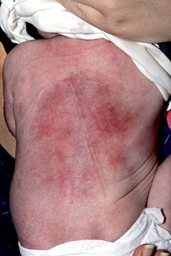
(BMJ)—A well 13-day-old boy presented w/ indurated, violaceous lesions on his back, along w/ hypercalcemia. The rash was initially treated as cellulitis, and he required IV fluids to correct the calcium. What was the dx?
Erysipelas
Sclerema neonatorum
Infantile hemangioma
Subcutaneous fat necrosis
Farber disease
You are correct. A clinical dx of subcutaneous fat necrosis was made after dermatology consultation. Subcutaneous fat necrosis is a rare, benign panniculitis occurring in full-term and post-term neonates that presents as skin-colored or erythematous nodules. Perinatal mechanical stress, tissue hypoxia, and hypothermia may contribute to pathogenesis. Skin bx can confirm dx. The condition is self-limiting, w/ a good prognosis; however, early recognition is important, as infants require monitoring for and correction of the associated hypercalcemia, thrombocytopenia, and hyperlipidemia. Severe hypercalcemia requires tx w/ IV fluids and furosemide, plus steroids and low-calcium feeds, if needed. This pt’s skin lesions and hypercalcemia resolved completely after 2mo.

BMJ 2018;363:k4062