(BMJ)—A 24-yo Filipino man presented w/ a 1-mo hx of a rapidly growing tender mass on his chest. Exam: vital signs WNL; 7x6-cm smooth, immobile, firm mass over upper sternum, w/ mild erythema, + cervical nodes. CT confirmed the dx. What is it?
Lipoma
Actinomycosis
Lymphoma
Tuberculosis
Chondrosarcoma
You are correct. This is a TB cold abscess of the chest wall due to hematogenous/lymphatic spread from active pulmonary TB. CT imaging showed the pulmonary TB was pleural based, and there was a separate large paraspinal TB abscess. A tuberculin skin test was positive at 48h. The quick growth of the chest wall mass in size and tenderness, w/ associated cervical lymphadenopathy, suggested TB, even though the pt was immunocompetent. TB was especially likely because he was originally from the Philippines, a TB-endemic area. The pt made a full recovery after tx w/ isoniazid, rifampicin, pyrazinamide, and ethambutol.

Emerg Med J 2018;35:586
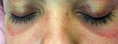
(BMJ)—A 34-yo woman presented w/ a 9-day hx of progressive bilateral limb weakness and mild dysphagia w/ fluids. Exam: periorbital rash. Grade 3-4/5 bilateral proximal limb and neck flexor weakness. Deep tendon reflexes and sensation intact. Labs: creatine kinase elevated; positive antinuclear matrix protein 2 antibody; weakly positive antinuclear antibody (1:80); negative anti-dsDNA antibody. MRI: hyperintensities in thigh muscles. What is the dx?
Thyrotoxic periodic paralysis
Systemic lupus erythematosus
Inclusion body myositis
Myasthenia gravis
Dermatomyositis
You are correct. The heliotrope rash and other typical findings (proximal limb weakness, mild dysphagia, preserved deep tendon reflexes, elevated creatine kinase, MRI findings) suggested dermatomyositis, and the dx was confirmed by muscle bx. In this pt, CT imaging of the chest, abdomen, and pelvis did not show any underlying malignancy, which is a concern in pts w/ dermatomyositis. She experienced substantial improvement of her weakness and dysphagia after high-dose steroids.

BMJ 2018;363:k3614
(BMJ)—An 11-yo boy w/ autism who ate only potato-containing foods presented w/ bilateral blurred vision, eye pain, and nyctalopia for several weeks. Exam: visual acuity 20/60 in both eyes; superficial punctate keratopathy; severe conjunctival xerosis. What is the dx?
Vitamin A deficiency
Episcleritis
Hypothyroidism
Vitamin E deficiency
Sjögren syndrome
You are correct. Vitamin A deficiency was confirmed by extremely low serum vitamin A levels. Conjunctival xerosis, a sign of long-standing vitamin A deficiency, is usually bilateral and reflects severe conjunctival dryness. In advanced cases, the entire conjunctiva may appear dry, roughened, thickened, corrugated, and sometimes skin-like. This pt’s ocular s/sx reversed following systemic vitamin A tx. As multiple nutritional deficiencies were evident, pediatrician and dietitian referrals were arranged.

Arch Dis Child 2018;103:890
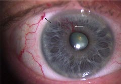
(BMJ)—A 73-yo man presented w/ a vascular lesion in his R eye. Exam: tortuous dilated vessel emerging from iris root to pupillary margin, w/ associated episcleral vessel. What is the dx?
Proliferative diabetic retinopathy
Arteriovenous malformation
Ocular ischemic syndrome
Uveitis
Retinal vein occlusion
You are correct. Further ophthalmic eval showed no evidence of posterior segment dz or underlying tumors, and a clinical dx of arteriovenous malformation was made. Neovascularization of the iris, also known as “rubeosis iridis,” results from retinal ischemia caused by proliferative diabetic retinopathy and retinal vein occlusion. Other causes for a vascular lesion of the iris include a dilated vessel from an underlying malignancy, and benign vascular tumors. Arteriovenous malformations are rare, nonprogressive vascular tumors that require an ophthalmic assessment to r/o other pathology.

BMJ 2018;362:k3699
By vgreene, 10 January, 2019