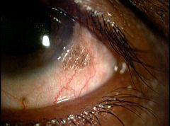
(BMJ)—A 35-yo alcoholic man presented w/ a lesion in his L eye. He had no visual problems and no signs of malnutrition or GI dz. Exam: L eye w/ triangular lesion in temporal interpalpebral bulbar conjunctiva. Labs confirmed the dx. What is it?
Conjunctival intraepithelial neoplasia
Bitot spot
Conjunctival nevus
Pterygium
Angular conjunctivitis
You are correct. Serum vitamin A level was low, confirming a Bitot spot assoc w/ vitamin A deficiency. Histopathologically it is characterized by a loss of goblet cells, accumulation of Gram-positive bacilli, and keratinization of the conjunctival epithelium. Most Bitot spots disappear after the pt receives systemic vitamin A, but up to a quarter may not respond completely. This pt received oral vitamin A (200,000 IU on days 1, 2, and 14) and the spot disappeared after 3mo.

BMJ 2016;355:i6032