(BMJ)—A 27-yo woman presented w/ painful leg lesions, despite 2wk of tx w/ amoxicillin/clavulanate for presumptive bilateral cellulitis. She had started an OCP 3mo prior. Exam: afebrile; tender, red, warm plaques w/ confluent palpable nodules on legs. Labs, UA, and CXR all normal. What is the dx?
Calcifying panniculitis
Nodular vasculitis
Thrombophlebitis
Drug-induced erythema nodosum
Pyoderma gangrenosum
You are correct. Bx confirmed erythema nodosum, a panniculitis subtype. Most cases are idiopathic, but frequent causes include infxn, sarcoidosis, vasculitis, connective tissue dz, and drugs (eg, abx, OCPs). Tx is supportive, w/ NSAIDs and topical steroids. Most cases spontaneously resolve. The pt stopped the OCP and was treated w/ topical steroids. After 2mo, the lesions completely disappeared, and no relapse occurred.

Emerg Med J 2017;34:194-197.
(BMJ)—An otherwise healthy 28-yo man presented w/ a 3-yr hx of gradually worsening visual acuity. Visual acuity exam: L eye (uncorrected), 20/100; L eye (corrected), 20/60. Scissoring of red reflex and conical reflection on nasal cornea when penlight shone from temporal side. What is the dx?
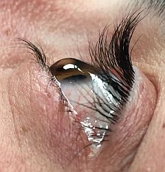
Fuchs corneal dystrophy
Terrien marginal corneal degeneration
Pellucid marginal degeneration
Keratoglobus
Keratoconus
You are correct. Keratoconus is a common corneal ectasia, a primary corneal dystrophy characterized by a change in thickness and curvature. Clinically, it presents as low visual acuity due to irregular astigmatism and increasing myopia. Cause is unclear, although risk factors include family hx, allergy, and repeated eye rubbing. This pt had implantation of intrastromal corneal ring segments, a well-tolerated and effective tx for pts w/ corneal ectasia (particularly keratoconus) that offers long-term improvement in visual, refractive, and keratometric measures.

BMJ 2017;357:j1461